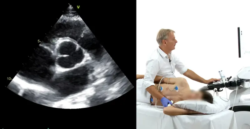
Do you know what you should look for on the echocardiography in patients with the referral “congenital defect”? Often these patients look fine and don’t know which defect they have.
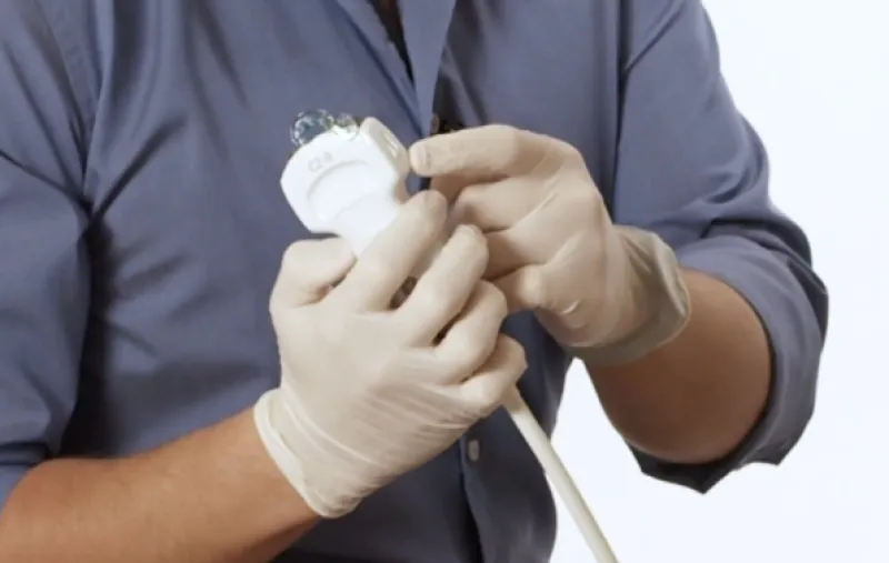
As sonographers, we try to get an appreciation of a 3D structure in the patient’s body using a 2D screen, which means our hand holding the transducer is constantly moving.
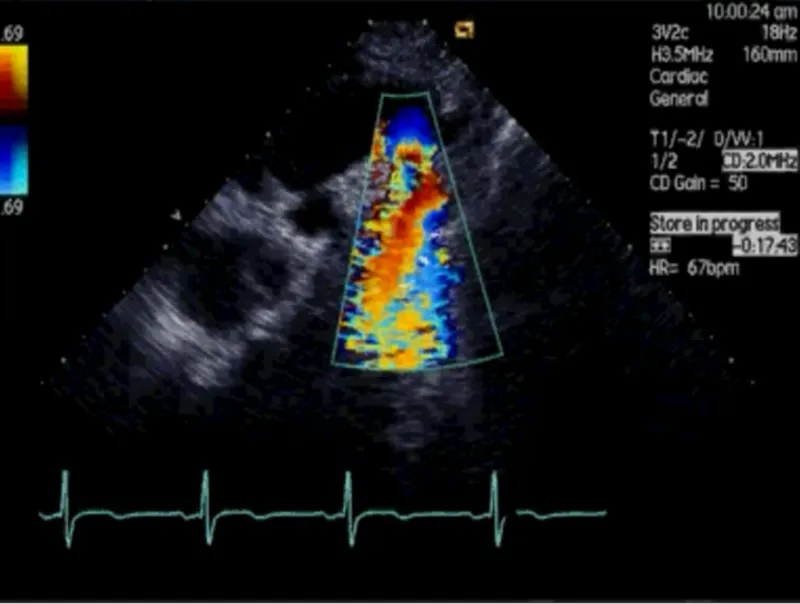
We all hate uncertainties and love numbers, which give us more guidance. This is the main reason the PISA method of quantifying mitral regurgitation appears so attractive. But should we use it in clinical practice...