
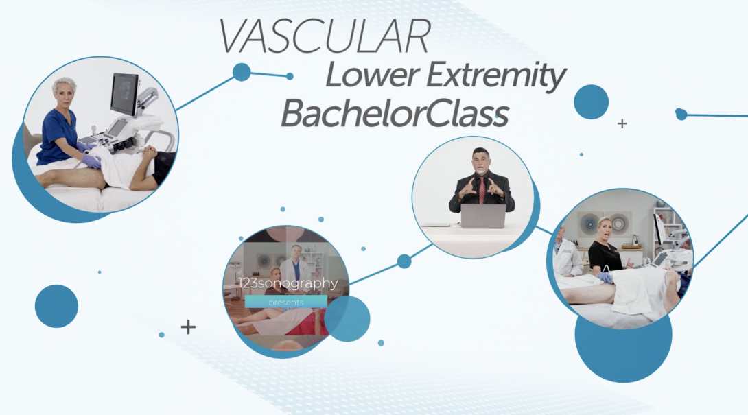


Course Speakers


Curriculum
Chapter 1
Free lectures
3 lectures and 0 quizzes
Lectures & Quizzes:
Chapter 2
1 CME
Basic and Advanced Ultrasound Concepts
In this first chapter we discuss the history of vascular disease, the importance of studying it, and its prevalence worldwide. Subsequently, we cover the professions to which this course is tailored and a chronological overview of the upcoming chapters. Prior to diving into the deep end of vascular imaging, we must first undersand the basic ultrasound concepts. Basic ultrasound concepts entail a general overview of the ultrasound system then a more focused lens on individual ultrasound system functions. Furthemore, we will learn how to set up our ultrasound environment. Once our ultrasound environment is prepared, we will review orientation, transducer ergonomics, transducer manipulation, and basic knobology. Then we proceed to learn advanced ultrasound concepts. These concepts entail image optimization via B-mode, color Doppler, and spectral Doppler, which will enable us to have full diagnostic confidence during image aquisition.
12 lectures and 1 quizzes
Lectures & Quizzes:
Chapter 3
0.5 CME
Clinical Ultrasound Concepts
A mastery of the technical and conceptual concepts regarding ultrasound imaging is not sufficient to create a competent sonographer. To truly excel, we must consider how we communicate with others. In order to answer the clinical questions, this chapter will provide a guideline regarding how communicate with both the patient and the physician.
2 lectures and 1 quizzes
Lectures & Quizzes:
Chapter 4
0.5 CME
Lower Extremity Arterial System
A thorough understanding of the peripheral arterial system is required to image the lower extremities. This chapter encompasses normal arterial anatomy, arterial physiology, and arterial hemodynamics.
3 lectures and 1 quizzes
Lectures & Quizzes:
Chapter 5
0.5 CME
Lower Extremity Arterial Pathology
Now that we have an understanding of normal peripheral arterial function, we have a foundation to learn how to differentiate between various peripheral arterial diseases. This chapter covers peripheral arterial pathology and the pathophysiology behind atherosclerosis, aneurysms, fibromuscular dysplasia, and congenital anomalies.
4 lectures and 1 quizzes
Lectures & Quizzes:
Chapter 6
0.75 CME
Lower Extremity Arterial Evaluation: Technical Protocol
This chapter covers the technical protocol for duplex evaluation of the lower extremity peripheral arterial system. We review how to orient via the anatomic landmarks. Subsequently, we review how to image the peripheral arterial system with B-mode, color Doppler, and sprectral pulsed wave Doppler.
13 lectures and 1 quizzes
Lectures & Quizzes:
Chapter 7
1.25 CME
Lower Extremity Arterial Evaluation: Abnormal Findings
This chapter covers duplex evaluation of lower extremity peripheral arterial pathology. We review how to image and document the pathological findings of stenosis, occlusion, and aneurysms via B-mode, color Doppler, and spectral Doppler.
14 lectures and 1 quizzes
Lectures & Quizzes:
Chapter 8
0.5 CME
Lower Extremity Venous System
A thorough understanding of the peripheral venous system is required to image the lower extremities. This chapter covers normal venous anatomy of the deep and superficial systems. Subsequently, this chapter covers venous physiology and venous hemodynamics.
3 lectures and 1 quizzes
Lectures & Quizzes:
Chapter 9
0.5 CME
Lower Extremity Venous Pathology
Now that we have an understanding of normal peripheral venous function, we have a foundation to learn how to differentiate between various peripheral venous diseases. This chapter covers peripheral venous pathology and the pathophysiology behind venous insufficiency, deep vein thrombosis, superficial thrombophlebitis, and post-thrombotic luminal wall changes.
3 lectures and 1 quizzes
Lectures & Quizzes:
Chapter 10
0.75 CME
Lower Extremity Venous Patency Evaluation: Technical Protocol
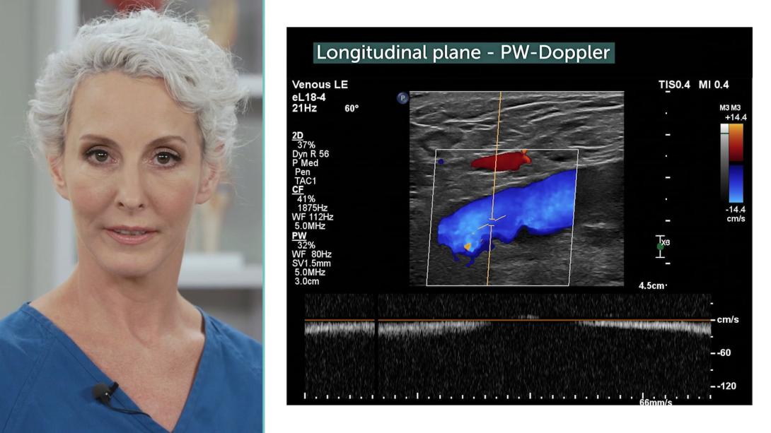
This chapter covers the technical protocol for duplex evaluation of the lower extremity peripheral venous system. We review how to orient via the anatomic landmarks. Subsequently, we review how to image the peripheral venous system for patency with B-mode, color Doppler, and sprectral pulsed wave Doppler.
10 lectures and 1 quizzes
Lectures & Quizzes:
Chapter 11
1.25 CME
Lower Extremity Venous Insufficiency Evaluation: Technical Protocol
This chapter covers the duplex evaluation of the lower extremity peripheral venous system. We review how to orient via the anatomic landmarks. Subsequently, we review how to image the peripheral venous system for insufficiency with B-mode, color Doppler, and spectral pulsed wave Doppler.
29 lectures and 1 quizzes
Lectures & Quizzes:
Chapter 12
1 CME
Lower Extremity Venous Evaluation: Abnormal Findings
This chapter covers the technical protocol for duplex evaluation of the lower extremity peripheral venous pathology. We review how to image and document the pathological findings of deep vein thrombosis, deep venous insufficiency, and superficial venous insufficiency via B-mode, color Doppler, and spectral Doppler.
15 lectures and 1 quizzes
Lectures & Quizzes:
Chapter 13
1 CME
Lower Extremity Functional Testing and Alternative Modalities
Functional arterial and venous testing is complementary to ultrasound imaging of the peripheral arterial and venous system. Together, these modalities allow us to paint a complete and vivid picture of the patient's peripheral arterial and venous pathology. This chapter covers ABI testing, segmental testing, and pulse-volume recording as well as air plethysmography testing. Furthermore, modern technology allows us to use alternative ultrasound modalities in order to improve our visualization of the peripheral venous system. This chapter also covers one of these modalities: intravascular ultrasound.
12 lectures and 1 quizzes
Lectures & Quizzes:
Objectives

After completing the Vascular Lower Extremity BachelorClass, you will be able to demonstrate scanning protocols for performing venous and arterial lower extremity vascular non-invasive ultrasound examinations.
Ideal for:

Student Discount
Are you a student? Get 50% discount on this course by completing the student application form.
Get Student DiscountPricing
One-Month Access
Take the most flexible route with a monthly subscription.
You get:
- Cancellation possible anytime after 4 months minimum run time
- Ability to complete quizzes and earn CME credits
Two-Year Access
If you want to take your time learning, this option is perfect for you. Save 40% on the 6-month option.
You get:
- 24 months access to our course
- Ability to complete quizzes and earn CME credits
One-Year Access
Our most recommended access duration to dive deep into the course. Save 30% on the 6-month option.
You get:
- 12 months access to our course
- Ability to complete quizzes and earn CME credits
Half-Year Access
Our shortest option for very fast learners. Ideal for people who have plenty of time to learn.
You get:
- 6 months access to our course
- Ability to complete quizzes and earn CME credits