1.6.2 Ultrasound Probe
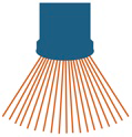
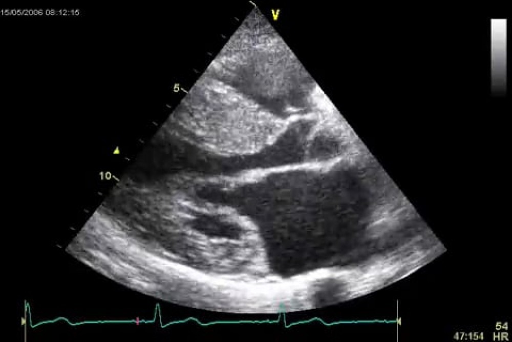
In echocardiography we use curvilinear probes. These have a small footprint (surface from which the ultrasound rays are emitted). Curvilinear probes emit the ultrasound beams in the form of a wave front. This is done by sequentially firing one element after the other. The ultrasound beam will move at an angle to the line of the piezoelectric array. Several such beams are used to create a sector which can be as wide as 90%. Such beams are emitted in rapid pulses and thus permit the creation of several sequential images as well as the demonstration of "motion".
The major advantage of this design is that the probes can be used to image even from small intercostal spaces. Therefore, ultrasound vendors take care to ensure that the "footprint" of the transducer is as small as possible. The ultrasound crystals emit a beam which has a certain width (beam width).
The width of the beam should be as narrow as possible. A narrow beam width improves image resolution. Usually the beam is focused on a certain region (i.e. near or far field). Like a camera this allows adjustment of the focus point. The more elements the transducer has (high scan line density) and the smaller the beam width is, the better is the "lateral" image resolution. The number of elements also influences the frame rate.
An important fact should be kept in mind: the smaller a certain sector is, the better is the resolution of the image because the processing function can be focused on a smaller region.
Scan line density is an important aspect of image quality because it permits higher image resolution.


