Scary Ultrasound – Spectacular images

Some of the things we see in ultrasound can be quite frightening. Since its Halloween I want to show you some pathologies that are quite spectacular. When showing these images (which were often incidental findings) lets not forget how important ultrasound was in detecting these specific pathologies. Ultimately, it helped to treat the underlying problem and often save lives.
So don’t be frightened
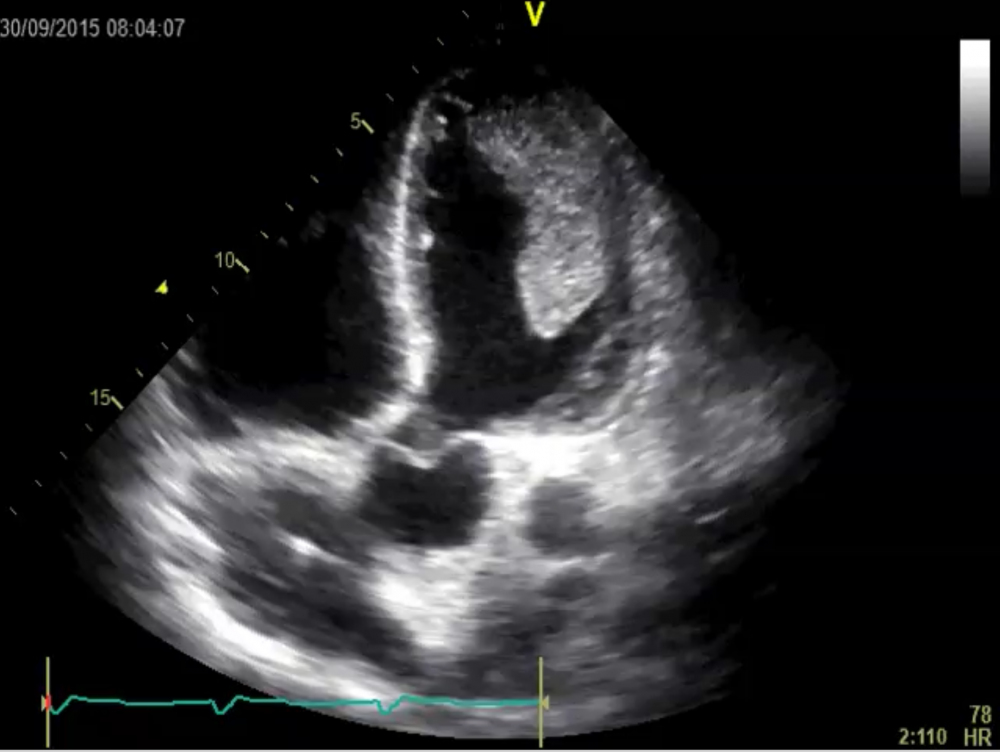
Gigantic thrombus in the left ventricle
This patient had an unnoticed large anterior myocardial infarction. He walked into our emergency department because of shortness of breath. As you can imagine we did not let him go home. The large thrombus resolved completely under warfarin therapy.
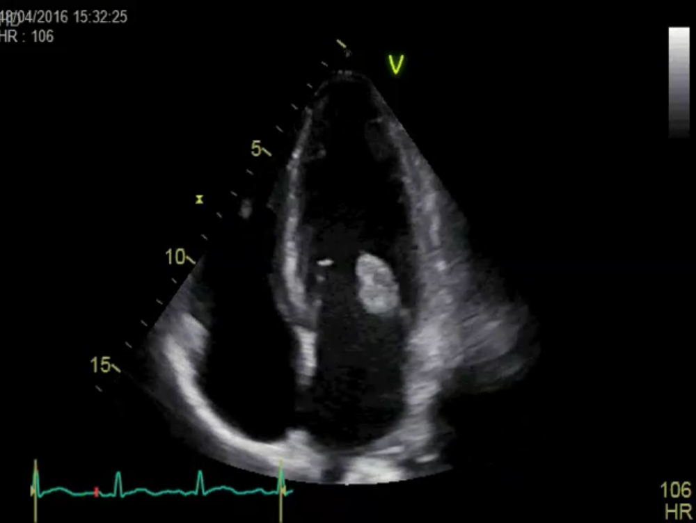
Highly mobile Vegetation on the MV
This patient had “fever of unknown origin” until an echo was performed. What is scary here is not only the size of the vegetation on the mitral valve but also its mobility. Believe it or not – the vegetation did not embolism! He was operated just in time!
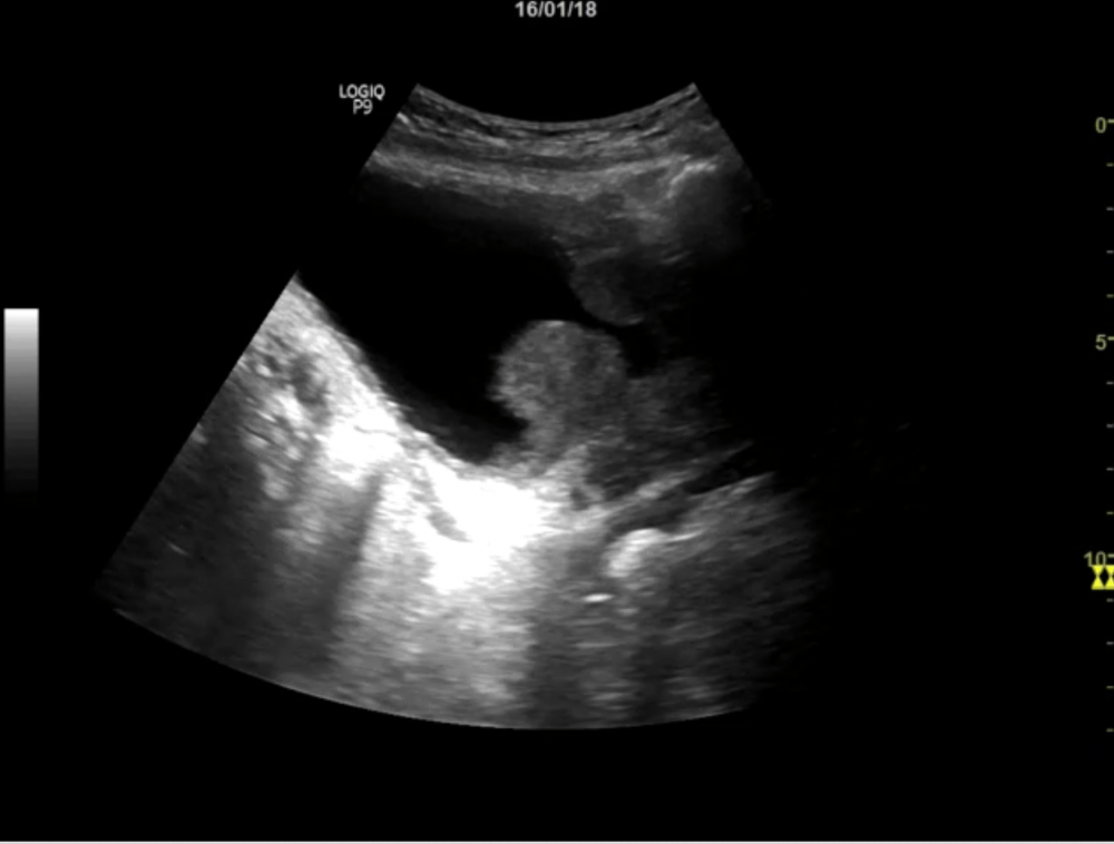
Huge Tumor of the Bladder
An elderly patient with hematuria and a bladder emptying disturbance. What we found was a huge carcinoma of the urinary bladder. The tumor invades the bladder wall and protrudes into the lumen.
Gastric outlet obstruction
Patient with alcohol abuse and recurrent duodenitis. The patient underwent Choledochojejunostomy, but again developed duodenitis. This led to stenosis of the pyloric region. One can see the massively enlarge fluid-filled stomach. The echo-free areas in the liver represent portocaval anastomosis (portal hypertension).
Aortic dissection and mobile thrombus
This patient had a peripheral embolism. When you see this image you will understand why. He not only has aortic dissection but also highly mobile vegetation on the dissection membrane.
ASD closure device “ping pong”
Do you see the ASD closure device bouncing back and forth in the left ventricle? This echo was performed one day after interventional ASD closure. Luckily the device was successfully retrieved and the patient did fine.
Prosthetic bypass graft
And finally here is a spooky vascular ultrasound of a prosthetic bypass graft, which has a chainsaw-like appearance when visualized via grayscale ultrasound. Due to the degree of invasiveness, this is only a final resort when treating a patient with severe arterial pathology. In this case, the patient had totally occluded stents in the superficial femoral artery. In order to bypass these occluded stents, the physician had to make an incision in the leg all the way from the common femoral artery to the above-knee popliteal artery."
For those of you who are looking for a Halloween treat, we have a special Halloween offer:
Don't let ultrasound frighten you! Benefit from extended access time - 12 months (instead of 6 months). Only until the 31st of October!


