
Where is Waldo (Part 1)?


This is the first of two blog posts in which I would like to “play” a similar game with you. I will call it the „where is the pathology“ game.
Let us see if you can find the “not so obvious” pathology „hidden“ in the following echo’s. The answers can be found at the end of this article.
Case 1 - Just an infarct?
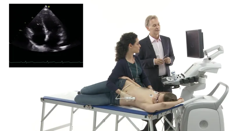
Here is the apical long axis view of a patient with an inferior myocardial infarction. The posterolateral wall is akinetic and the left atrium is enlarged. But, is there something else? (click images to see videos)

Case 2 - Mild aortic regurgitation so what?
What is hidden in this color Doppler study of an asymptomatic patient with a systolic murmur?

Case 3 – Don’t miss the problem here!

Unraveling the mysteries
Are you ready for the answers? If you did find “Waldo” congratulations, since detecting the pathology was highly relevant in all patients. It changed the management in all cases.
Organic mitral regurgitation
Case 1 shows a flail anterior leaflet. You have to look closely to see the flail portion of the leaflet, which protrudes into the left atrium. Here is another view that focuses on the mitral valve.

Mitral regurgitation in this patient was severe. Since he was symptomatic and because mitral regurgitation was organic and not functional he was sent to surgery for mitral valve repair.
Turbulent flow
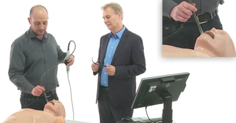
The key finding in case two is the turbulent flow in the left ventricular outflow tract. Elevated flow velocities in the LVOT can be caused either by dynamic (SAM-phenomena in hypertrophic cardiomyopathy) or fixed outflow tract obstruction. To clarify the pathology we performed a transesophageal study:

This patient has subvalvular (membranous) aortic stenosis. This explains both the systolic murmur and aortic regurgitation. The systolic high velocity jet is directed towards the aortic valve and causes damage to the aortic valve and regurgitation. The membrane was removed surgically.
The answer is not in the color

Clearly, there is a thrombus attached to the occluder. No wonder the patient had a minor stroke. The patient had to undergo operation with removal of the thrombus and the occluder.
Often we are too focused on the obvious and don’t look for the unexpected. In the first case we might think that mitral regurgitation was functional, caused by the inferior infarct. In the second case one might think turbulent flow is caused by valvular aortic stenosis, and in case three we are looking for a residual patent foramen ovale instead of looking at the morphology of the interatrial septum.
If you want to be an expert on finding the “Waldo’s” on the echocardiogram you might want to check out our echocardiography courses!
In any case, look out for the second part of “Finding Waldo”.
Recommended articles:
Where is Waldo on the Echo (Part 2)?
The View from the Top
Old and New
Related Courses
The Echo MasterClass is the most comprehensive premium online course in echocardiography,…
The Echo BachelorClass provides an invaluable foundation for the practice of echocardiography…
The TEE MasterClass is an exclusive teaching program that will allow you to learn the art of…