
Icing on the cake

Our blog p osts published thus far have mainly dealt with practical issues that assist you in learning the tips and tricks of echocardiography. From your feedback we gather that some of you are far more advanced and ready for some high-end echo techniques. Don’t worry: we will not abandon our bread-and-butter strategy, but will merely add a little icing to the cake.

One technique, which is certainly becoming more widespread as time goes by, is 3D echocardiography. Is 3D really helpful? Will it replace 2D? In what situations should it be used?
Read this case and judge for yourself ...
A 23-year-old woman from the Ukraine was referred to us for evaluation of an atrial shunt. Specifically, we had to determine whether interventional closure was feasible.
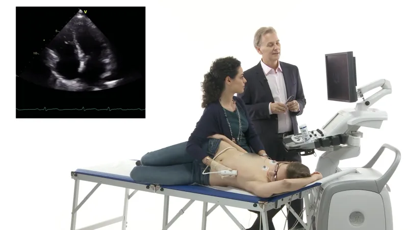
 Four-chamber view showing a large right ventricle.
One thing was very obvious on this transthoracic echo: the atrial septal defect that had been diagnosed several years ago was associated with a large shunt. Look how large the right ventricle is. Its diameter is 48 mm - in a rather small young woman.
 Color Doppler study of an atypical short axis view (color Doppler).
Atrial shunts are usually seen best on atypical views as the one above. This picture shows a rather broad jet from the left atrium into the right one. The defect, it would appear, is really very large. A large secundum atrial septal defect. Is it too large to be closed by using an interventional approach? We were told, from previous studies, that the defect was fenestrated. The question is: what does the rim of the defect look like? How much residual tissue is left? To answer this question we had to perform a transesophageal study:
 TEE shows a large secundum ASD measuring almost 24 mm. Still, it looked as if there was residual tissue left.
We performed a 3D study as well. This is what we saw:
 3D TEE study of the atrial septal defect. Note that it is oval inshape and there is a septation which divides the defect into a smaller and a larger portion.
The 3D study clearly showed that the defect was large and oval, but was not fenestrated. It merely had a septation. There was no place to hold the occluder. We decided that interventional occlusion was not possible. Closure would have to be performed by conventional surgery. Back to our question: Is 3D better than 2D? My take on this issue: no, but it can provide additional information. It is the icing on the cake of 2D imaging. It may certainly help in the management of patients. Atrial septal defects are just one example.
Watch out for more practical examples of 3D echo and also other new technologies such as speckle-tracking and deformation imaging in one of our upcoming posts.
If you have not already done so, check out our Masterclass. The Masterclass will teach you everything you need to know about transthoracic echo in just 90 days. Click here!
Related Courses
The Echo BachelorClass provides an invaluable foundation for the practice of echocardiography…